


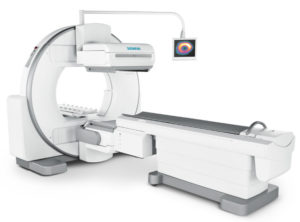
Partners offers Nuclear imaging with a Siemens Excel Gamma Camera
These medical imaging modalities are used to gather functional details about the body. Gamma studies are physiological tests and differ from MRI and CT scans, which are largely anatomical imaging tools. For example, cardiac stress tests, gastric emptying, and triple phase bone scans are studies that give us functional information about how the body is working and would need to be performed on a Gamma Camera.
 Gamma Imaging works by ingesting or injecting the patient with a radiopharmaceutical which is targeted to work on specific parts of the body. Patients then wait for a period of time while the radiopharmaceutical is absorbed, uptake times can range from minutes to hours depending on the type of study ordered.
During the study, the patient rests comfortably on a table while two or three imaging detectors move around and gather information on how their body is functioning.
After imaging, the machine creates a one, two or three-dimensional image of the study and provides the necessary information for the radiologist to make a diagnosis.
Common gamma studies performed at Partners Imaging Centers are triple phase bone scans, cancer imaging bone scans, liver/spleen scans, brain SPECT and gastric emptying studies.
Always ask your doctor about having your nuclear medicine procedure performed at Partners Imaging compared to larger inpatient facilities. In most cases Partners Imaging is less expensive and can be scheduled almost immediately.