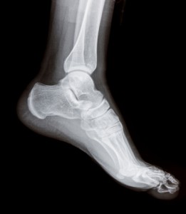
Partners Imaging have two digital, low radiation, U arm X-Ray machines for patients on a walk-in basis
These are generally quick scans to perform and patient comfort is optimal on a walk-in basis. How it works – Radiographs are produced by the transmission of x-rays through a patient to a capture device then converted into an image for diagnosis. In Film-Screen radiography an x-ray tube generates a beam of x-rays which is aimed at the patient. The x-rays which pass through the patient are filtered to reduce scatter and noise and then strike an undeveloped film, held tight to a screen of light emitting phosphors in a light-tight cassette. The film is then developed chemically and an image appears on the film.
 Diagnostic x-ray technology was discovered in 1895 by a German physicist, Wilhelm Roentgen. His discovery precipitated one of the most important medical advancements in human history. X-rays lets physicians see straight through human tissue to examine bones, cavities and other soft tissue (lungs, blood vessels).
In radiology today most imaging modalities are digital. MR, CT, PET, and ultrasound, all offer high-resolution digital images that can be shared and distributed widely. Digital X-Ray now allows Partners Imaging to provide one of the most common types of basic imaging exams in digital format, making routine X-Ray more efficient and convenient for your Physicians.
Partners Imaging uses the latest U-Arm technology. This allows the patient to stand and the X-Ray is moved into position for the best image. These X-Rays use a low radiation digital detector for high quality images. This allows us to perform general x-ray while capturing all of the clinical data on computers instead of film. Radiologists can now view the studies on a computer screen, giving them the ability to enhance images or change contrast for better diagnostic capabilities, and share them with other Physicians via the Partners PACS system. The digital system also reduces patient exposure to excessive x-rays by cutting down on repeat exams because of poor film quality or patient movement.


